
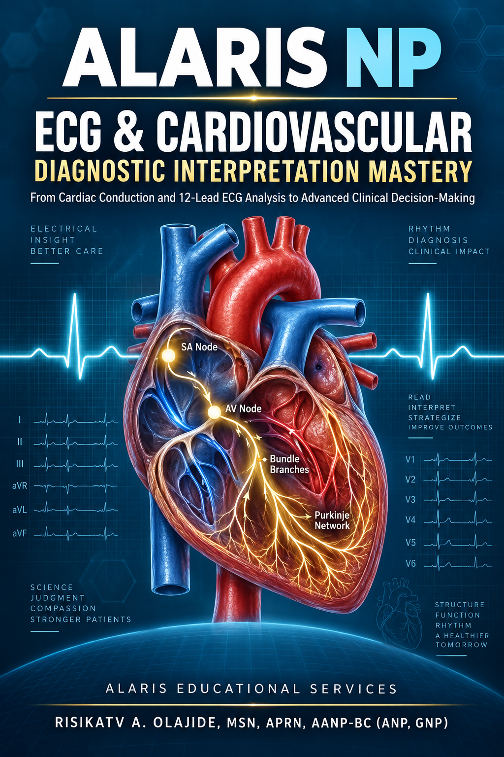
Description
BUILD CONFIDENCE. RECOGNIZE THE PATTERN. CONNECT THE FINDINGS. MAKE THE CLINICAL DECISION.
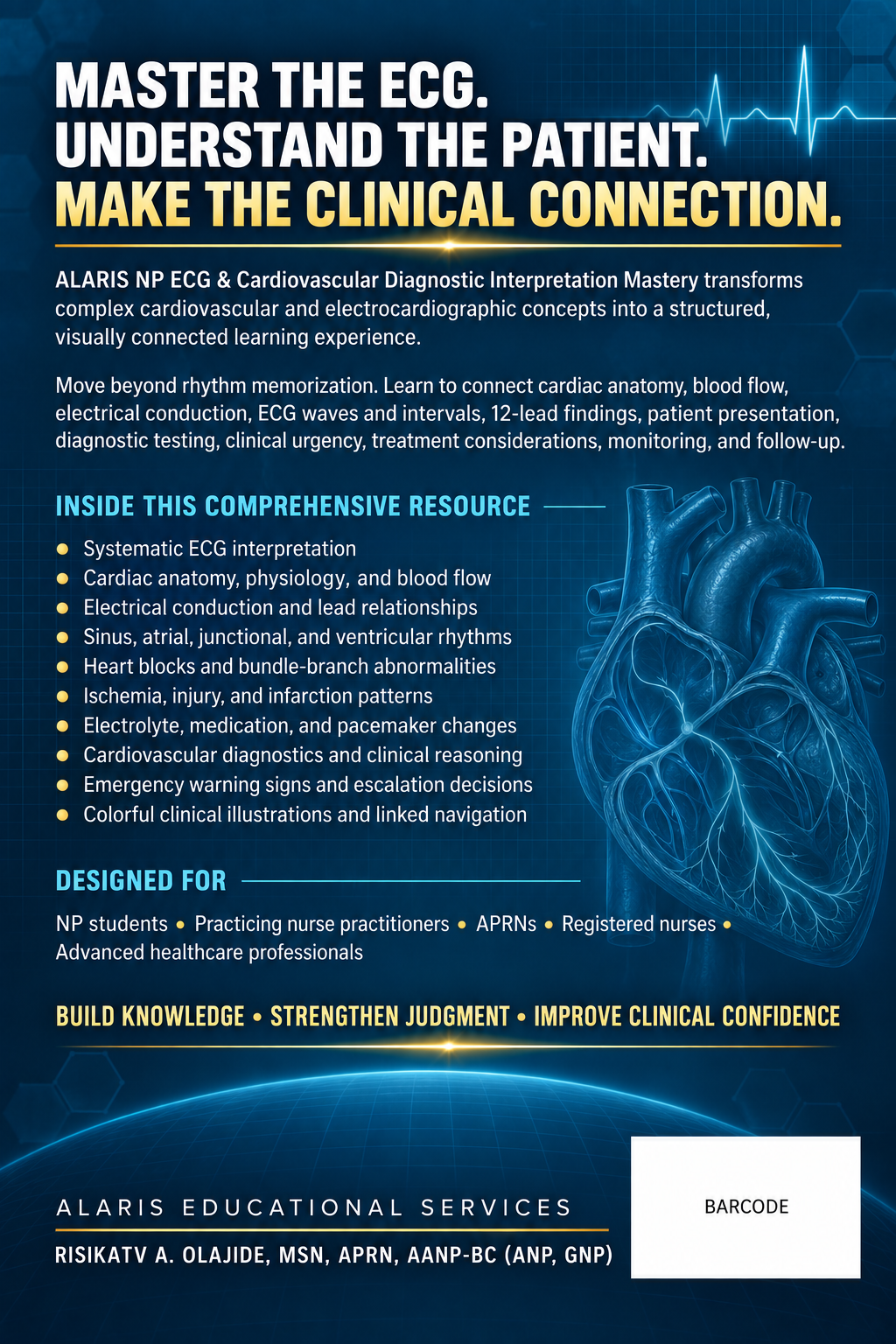
ALARIS NP ECG & Cardiovascular Diagnostic Interpretation Mastery is a comprehensive, visually integrated educational resource created to help nurse practitioner students, practicing nurse practitioners, APRNs, registered nurses, and other advanced healthcare professionals understand ECG interpretation within the complete cardiovascular clinical picture.
This is more than an ECG rhythm-recognition guide.
The resource teaches learners how to connect cardiovascular anatomy, blood flow, cardiac electrical conduction, ECG waves and intervals, lead findings, patient symptoms, physical examination findings, diagnostic-test results, medications, clinical urgency, and management decisions.
Instead of relying only on memorization, students are guided through a systematic process for determining:
• What the tracing demonstrates
• Which cardiac structure or electrical pathway is involved
• Whether the finding may be normal, abnormal, or immediately dangerous
• Which clinical conditions should be considered
• What additional information or diagnostic testing is needed
• Which findings require emergency intervention or specialty referral
• How treatment, monitoring, patient education, and follow-up may be approached
A SYSTEMATIC APPROACH TO ECG INTERPRETATION
Learners are taught to evaluate ECGs in an organized sequence, including:
• Rhythm regularity
• Atrial and ventricular rates
• P-wave morphology and atrial activity
• PR interval and atrioventricular conduction
• QRS width, morphology, and ventricular depolarization
• Cardiac axis
• R-wave progression
• ST segments
• T-wave morphology
• QT and corrected QT intervals
• Abnormal Q waves
• Lead-to-lead relationships
• Reciprocal and contiguous-lead changes
• Comparison with previous or serial ECGs
• Correlation with symptoms, vital signs, laboratory results, and clinical stability
COMPREHENSIVE CARDIOVASCULAR CONTENT
The resource provides extensive instruction across major ECG and cardiovascular areas, including:
• Cardiovascular anatomy and physiology
• Circulation through the heart, lungs, coronary vessels, and systemic circulation
• Cardiac chambers, valves, major vessels, and coronary circulation
• Cardiac action potentials and impulse formation
• Sinoatrial, atrioventricular, His-Purkinje, bundle-branch, and ventricular conduction
• ECG paper, calibration, measurements, and technical principles
• Correct electrode positioning and 12-lead ECG placement
• Limb leads, augmented leads, and precordial leads
• Electrical vectors, cardiac axis, and lead orientation
• Normal sinus rhythm and sinus-node abnormalities
• Premature atrial, junctional, and ventricular complexes
• Atrial fibrillation and atrial flutter
• Supraventricular tachycardias
• Junctional rhythms
• Ventricular tachycardia and ventricular fibrillation
• Atrioventricular conduction blocks
• Right and left bundle-branch blocks
• Fascicular blocks and intraventricular conduction abnormalities
• Bradycardias and tachyarrhythmias
• Chamber enlargement and ventricular hypertrophy
• Myocardial ischemia, injury, and infarction patterns
• ST-segment and T-wave abnormalities
• QT prolongation and associated dysrhythmia risk
• Electrolyte- and metabolic-related ECG changes
• Medication-related ECG abnormalities
• Pacemaker rhythms and common pacemaker concerns
• Cardiomyopathies, heart failure, valvular disorders, and pericardial conditions
• Hypertension, coronary artery disease, and acute coronary syndromes
• Syncope, palpitations, dyspnea, chest pain, and hemodynamic instability
• Cardiovascular diagnostic testing and interpretation
• Emergency cardiovascular recognition and escalation of care
COLORFUL CLINICAL ILLUSTRATIONS THAT CONNECT THE CONCEPTS
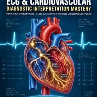
Colorful, clinically focused illustrations and teaching diagrams help students visualize how the cardiovascular system functions.
The visual instruction connects:
• Blood flow through the heart, lungs, and body
• Heart chambers and valves
• Coronary arteries and myocardial blood supply
• Electrical impulse formation and conduction pathways
• Depolarization and repolarization
• P waves, QRS complexes, ST segments, T waves, and intervals
• Lead placement and the areas of the heart viewed by each lead
• Conduction abnormalities and their ECG manifestations
• Cardiovascular disease processes and related clinical findings
Arrows, labels, color-coded pathways, comparison diagrams, and ECG examples help transform difficult concepts into understandable clinical relationships. The illustrations are integrated into the teaching material rather than included only as decoration.
CLINICAL REASONING AND DECISION-MAKING
ECG findings are interpreted in the context of the whole patient. Learners are encouraged to connect each tracing with:
• Presenting symptoms
• Relevant history and cardiovascular risk factors
• Vital signs and hemodynamic stability
• Focused cardiovascular examination findings
• Differential diagnoses
• Laboratory and biomarker results
• Imaging and diagnostic studies
• Medication history
• Potential adverse drug effects
• Immediate safety concerns
• Appropriate treatment priorities
• Referral and escalation decisions
• Monitoring and reassessment
• Patient and family education
• Follow-up and prevention
The resource emphasizes the critical principle that an ECG should not be interpreted in isolation. A tracing must be evaluated together with the patient’s symptoms, examination findings, risk factors, diagnostic results, and overall clinical condition.
DESIGNED FOR EFFICIENT NAVIGATION
The organized heading structure and linked Table of Contents help students move directly to the information they need.
Students can use the navigation system to:
• Select a chapter or major cardiovascular topic from the Table of Contents
• Move directly to specific rhythms, conduction abnormalities, diagnostic findings, or clinical conditions
• Locate focused explanations without searching through the entire resource
• Review a difficult topic before class, simulation, clinical rotation, or examination
• Compare related rhythms and cardiovascular abnormalities
• Return to foundational anatomy, blood flow, or conduction concepts when clarification is needed
• Use the resource as both a structured learning program and a rapid clinical-reference tool
The learner may study the book from beginning to end or move directly to a selected topic using the linked navigation.
WHO SHOULD USE THIS RESOURCE?
This educational resource is designed for:
• Family Nurse Practitioner students
• Adult-Gerontology Primary Care NP students
• Adult-Gerontology Acute Care NP students
• Emergency and urgent-care NP students and clinicians
• Psychiatric NPs seeking stronger medical assessment skills
• Practicing nurse practitioners
• APRNs and registered nurses
• Graduate nursing students
• Clinicians reviewing cardiovascular assessment and ECG interpretation
• Learners preparing for examinations, clinical simulations, rotations, or professional practice
HOW THIS RESOURCE CAN SUPPORT STUDENTS
Students can use this resource to:
• Develop a consistent ECG interpretation method
• Build a stronger foundation in cardiovascular anatomy and physiology
• Understand how electrical activity creates ECG waveforms
• Recognize common and dangerous rhythm abnormalities
• Differentiate similar-looking ECG patterns
• Connect ECG findings with patient presentations
• Identify red flags and time-sensitive cardiovascular emergencies
• Strengthen cardiovascular differential diagnosis
• Improve diagnostic-test interpretation
• Review pharmacologic and nonpharmacologic considerations
• Prepare for clinical rotations and simulation experiences
• Reinforce assessment, diagnosis, planning, intervention, and evaluation skills
• Support board-review preparation and continued professional development
• Improve clinical communication and documentation of cardiovascular findings
WHAT MAKES THIS RESOURCE DIFFERENT?
ALARIS NP ECG & Cardiovascular Diagnostic Interpretation Mastery combines foundational science, ECG interpretation, cardiovascular diagnostics, clinical reasoning, emergency recognition, treatment considerations, patient education, and follow-up within one integrated learning resource.
It is designed to help learners move beyond asking, “What rhythm is this?” and begin asking:
“What does this finding mean for this patient, how urgent is it, what additional information is needed, and what should happen next?”
Published by: ALARIS EDUCATIONAL SERVICES
Author: RISIKATV A. OLAJIDE, MSN, APRN, AANP-BC (ANP, GNP)
Reviews
There are no reviews yet.